You can kill Covid with a flick of a switch, study shows (www.israel21c.org)
Tel Aviv research: 99.9% of COVID-19 virus dead in 30 seconds with UV LEDs (www.jpost.com)
Cheaper LEDs can disinfect against COVID-19, Israeli scientists find (www.timesofisrael.com)
These are the first half-dozen of over 200 online articles that were over a period of approximately two weeks following the publication of this Tel Aviv University press release, dated December 14, 2020:
with the subtitle, “Groundbreaking research finds UV-LED diodes efficiently and cheaply disinfect social spaces.”
“Groundbreaking” research? This has a touch of hyperbole, but let’s see …
UV-LED Disinfection
On September 10, 2020, the respected Journal of Photochemistry & Photobiology, B: Biology published this paper:
Gerchman, Y., et al. 2020. “UV-LED disinfection of Coronavirus: Wavelength effect,” J. Photochemistry & Photobiology B: Biology 212 (2020) 112044 (DOI: 10.1016/j.photobiol.2020.112044).
The paper is open-access, for which the publisher deserves due credit for making its COVID-19-related research papers freely available.
The paper’s abstract is interesting:
“UV light-emitting diodes (UV LEDs) are an emerging technology and a UV source for pathogen inactivation, however low UV-LED wavelengths are costly and have low fluence rate. Our results suggest that the sensitivity of human Coronavirus (HCoV-OC43 used as SARS-CoV-2 surrogate) was wavelength dependent with 267 nm ~ 279 nm > 286 nm > 297 nm. Other viruses showed similar results, suggesting UV LED with peak emission at ~286 nm could serve as an effective tool in the fight against human Coronaviruses.”
but the introduction is more informative:
“Numerous studies have examined the sensitivity of different microorganisms (including viruses) to UV LED at different wavelengths as detailed in Table 1, for suspended viruses. However, no study to date has examined the efficiency [sic] of UV LEDs at different wavelengths on the inactivation of the human corona virus. Here, we have used the human coronavirus OC43 (HCoV-OC43) as a surrogate to the SARS-CoV-2, to develop a dose-response curve for UV-LEDs at various wavelengths.”
.. and here we need to pause in order to put these statements into context. The authors referenced twelve previous studies in their Table 1, but the key phrase here is “UV LED.” If we generalize this to “ultraviolet (UV) radiation,” there are many more studies of the relationship between wavelength and the efficacy (not “efficiency”) of UV radiation in inactivating viruses. In fact, the first study was published 144 years ago (Downes and Blunt 1877). The virucidal action spectrum for UV radiation was first established by Rivers and Gates (1928) and Sturm et al. (1932).
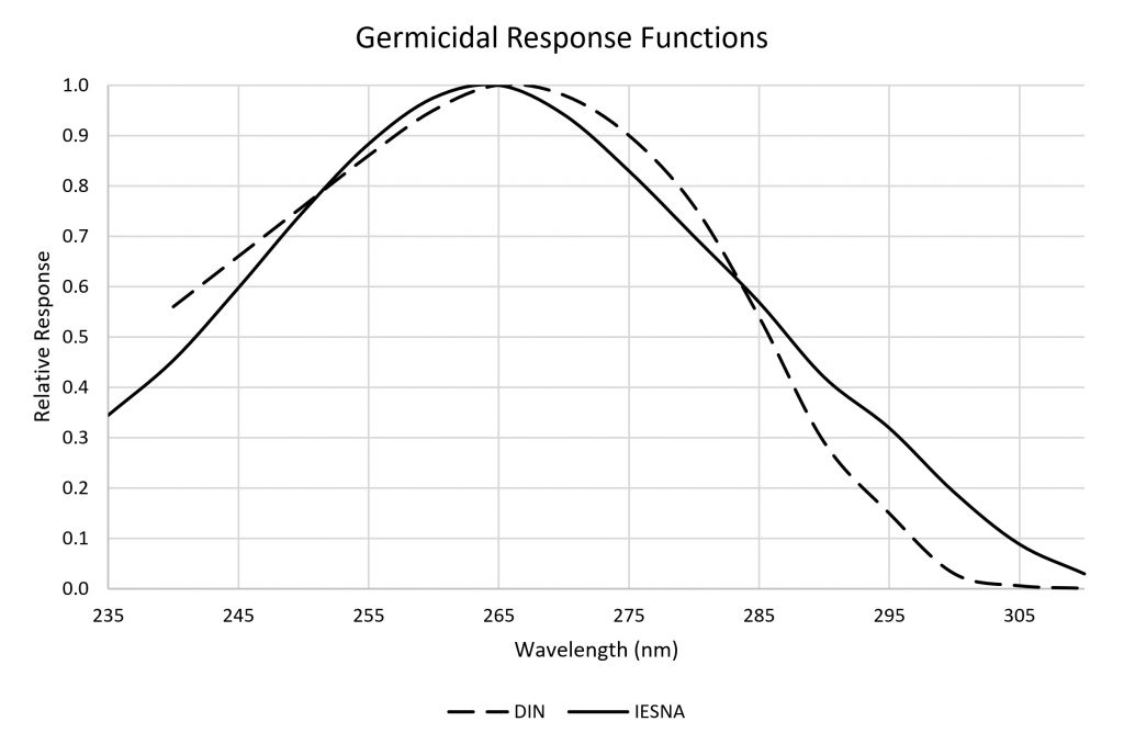
Figure 1 shows the action spectra (DIN and IES) for germicidal ultraviolet radiation applications, such as upper-room air and municipal water disinfection, that have been widely adopted by the CIE, IES, ACGIH, NIOSH, DIN, and other standards organizations:
FIG. 1 – Standard germicidal response functions.
What is rarely mentioned is that these action spectra are based on laboratory results with the Escherichia coli bacterium (e.g., Gates 1930). It is not a coincidence that the peak response near 265 nm corresponds with the peak spectral absorptance of deoxyribose nucleic acid (DNA) – UV radiation disrupts the genetic code of viruses, bacteria, and fungi, thereby preventing them from reproducing (e.g., Hollaender and Oliphant 1944).
The peak spectral response of different viruses and other pathogens may therefore vary by perhaps five nanometers or so (e.g., Linden 2001). However, the DIN and IES action spectra remain applicable for practical applications of germicidal UV radiation.
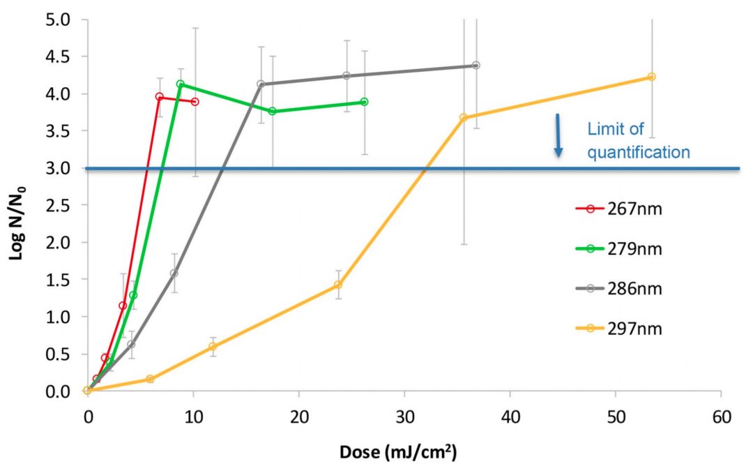
… which brings us back to the current paper of Gerchman et al. (2020). The results presented in the paper are summarized in Figure 2, where the dose refers to the UV irradiance multiplied by the exposure time, and the horizontal “limit of quantification” line represents the dose required to achieve log-three (99.9 percent) inactivation of the virus colony:
We may compare this to the IESNA germicidal response curve as enumerated in CIE 155:2003, Ultraviolet Air Disinfection, relative to the peak response at 265 nm:
Wavelength
Gerchman et al.
IESNA
267 nm
1.00 ± 0.09
0.990
279 nm
0.82 ± 0.03
0.726
286 nm
0.46 ± 0.05
0.543
297 nm
0.19 ± 0.05
0.268
Table 1 – Spectral response comparison.
The error bars shown in Figure 2 represent standard deviations in the results, which the authors explained as “an artifact due to lack of precision in enumerating the low number of [virus] survivors.” It is refreshingly surprising to see such honesty in published results; this sort of information is usually confined to supplementary material so as to not “confuse” the reader with data that might weaken the paper’s conclusions.
Whether there are errors of fact or the authors used questionable experimental procedures is not a topic that I as a science journalist (among other things) am academically qualified to comment on. What I will say, however, is that the paper itself has been carefully constructed and well-written, and is a model of academic writing.
So far, so good. However, there are no surprises here – the OC43 coronavirus appears to be somewhat less susceptible to longer wavelengths than E. coli bacteria, but this does not invalidate the applicability of germicidal response functions shown in Figure 1. The research is if anything no more than a confirmation of accepted scientific fact – viruses are susceptible to ultraviolet radiation, with a peak response near the DNA peak spectral absorptance of 265 nm.
The question then is, why did the editors of the Journal of Photochemistry & Photobiology decide that the paper was worthy of publication? While it is impossible to speak on behalf of the editors, one answer is that there is often value in the publication of negative results.
If the research had shown that the OC43 coronavirus was highly susceptible to longer wavelengths of UV radiation, that would have been stunning news that would have more than justified publication of the paper. Instead, the study merely confirmed that the existing standard germicidal response curves are generally applicable to HCoV-OC43, and (presumably) to SARS-CoV-2.
The academic value of the paper is therefore in describing what appears to be a carefully designed and executed series of experiments that yielded negative results. It informs other researchers of what has been done, and so allows them to direct their research efforts elsewhere.
Media Relations
There have to date been over 54 thousand academic papers relating to the SARS-CoV-2 virus that have been published in biomedical and life science journals. The probability of any one article coming to the attention of the public is basically infinitesimal … if it were not for university media relations officers.
The role of the media relations officer is to present the often-arcane details of academic research to the public. For both public and private universities, being seen in positive terms by the public is key to obtaining financial support from both public and private institutions. In other words, the role of a media relations officer is that of a marketing professional.
It is often a difficult job – how do you take a paper with a random title such as, “Genetic diversity of the Plasmodium falciparum GTP-cyclohydrolase 1, dihydrofolate reductase and dihydropteroate synthetase genes reveals new insights into sulfadoxine-pyrimethamine antimalarial drug resistance” (DOI:10.1371/journal.pgen.1009268), and present it to a public more interested in YouTube celebrities and sports figures? You begin your press release with a catchy title such as, “New mutations in malaria parasite encourage resistance against key preventative drug,” but the only qualification for the job is usually a bachelor’s degree in journalism. The media relations officer, through misunderstanding their interview with the researcher or lack of knowledge and experience, may fail, sometimes spectacularly.
Tel Aviv University
I have no desire to name and shame the media relation officer at Tel Aviv University in Israel, but I must say something. Here are a few select quotes for the aforementioned press release of December 14th, 2020:
“Researchers from Tel Aviv University have proven that the coronavirus can be killed efficiently, quickly and cheaply using ultraviolet (UV) light-emitting diodes (UV-LEDs). This is the first study in the world conducted on the disinfection efficiency of a virus from the family of coronaviruses using UV-LED irradiation at different wavelengths or frequencies.”
This may be technically true, but the significance of this study is grossly overrated in calling it, “the first study in the world.”
“In the study, the researchers tested the optimal wavelength for killing the coronavirus, and found that a length of 285 nanometers was almost as efficient in disinfecting the virus as a wavelength of 265 nanometers, requiring less than half a minute to destroy more than 99.9% of the coronaviruses.”
It does not take a mathematician to understand that a relative efficacy of 46 ± 5 percent is not “almost as efficient.”
“The entire world is currently looking for effective solutions to disinfect the coronavirus … The disinfection systems based on LED bulbs, however, can be installed in the ventilation system and air conditioner, for example, and sterilize the air sucked in and then emitted into the room.”
This is true, but it has nothing to do with the research paper. The popular press has been awash with stories about germicidal disinfection using radiation, a technology that has been in commercial use since 1909 (von Recklinghausen 1914). Economics currently favour low-pressure mercury vapour lamps that emit ultraviolet-C radiation at 254 nm, but rapid progress is being made in the development of more efficient and inexpensive ultraviolet LEDs. Again, nothing to do with this paper.
Quoting one of the paper’s authors from the press release, “We discovered that it is quite simple to kill the coronavirus using LED bulbs that radiate ultraviolet light, but no less important, we killed the viruses using cheaper and more readily available LED bulbs, which consume little energy and do not contain mercury like regular bulbs.”
This is … painful. Even if the author meant specifically the OC43 coronavirus and ultraviolet radiation generated by light-emitting diodes rather than mercury-vapour lamps, stating that anything was “discovered” is simply ludicrous.
It should further be noted that the radiant efficacy of commercially-available 285 nm UV-LEDs is currently on the order of one percent. This may be compared to that of mercury-vapour lamps, with efficacies on the order of 40 percent. The advantage of UV-LEDs is that it is much easier to direct their emitted radiation into narrow beams, a requirement for upper-room air disinfection devices.
Finally:
“Last year, a team of researchers led by Prof. Mamane and Prof. Gerchman patented a combination of different UV frequencies that cause dual-system damage to the genetic load and proteins of bacteria and viruses, from which they cannot recover – which is a key factor that is ignored.”
This is one of those, “Wait, what?” moments, where an entirely different and much more relevant press release could have been written about this technology. The patent application in question is US Patent Application 20200255305, “Method and Device for Water Disinfection,” that uses two sources to simultaneously emit UV-C and UV-B radiation. Patent applications are not peer-reviewed, however, and so the information needs independent verification and much more detail concerning the experiments behind the invention.
The primary problem is that the media relations officer was clearly struggling to understand the issues and write a coherent and informative press release. At best, there appears to have been “a failure to communicate.”
Viral Misinformation
Whatever misinformation and confusion there may have been in the press release, it won the lottery in being selected by the mass media for amplification. Looking at only the first six of some 200 article titles:
“99.9% of Covid-19 virus dead in 30 seconds with UV LEDs, says Tel Aviv research.”
No, neither the paper nor the press release made any claims regarding the SARS-CoV-2 virus that causes the COVID-19 disease. It explicitly stated that the HCoV-OC43 virus (which is one of the many viruses responsible for the common cold) was chosen as a surrogate for the SARS-CoV-2 virus, and that “… our future work will confirm these results by testing the impact of LEDs and their combinations on SARS-CoV-2.”
“You can kill Covid with a flick of a switch, study shows.”
The paper never suggested such an outlandish idea. It instead focused on the relative dose of ultraviolet radiation at different wavelengths to inactivate (not “kill”) a particular virus. The irradiance levels employed in the experiments would be totally impractical for surface disinfection in the real world.
“Tel Aviv research: 99.9% of COVID-19 virus dead in 30 seconds with UV LEDs.”
The paper uses the noun “seconds” just once, referring to “… up to 60 s for 267 and 279 nm and up to 90 s for 286 and 297 nm.” The key metric is dose – ultraviolet irradiance multiplied by exposure time. Whoever wrote this headline simply invented the number as clickbait.
… and so it goes, like some sinister version of the children’s game Chinese whispers. Perfectly reasonable and valid scientific information is endlessly repeated and distorted from paper to press release to mass media articles. Like most such events, the story will have a half-life measured in weeks to a few months before it is forgotten. Unfortunately, the misinformation spreads like a virus, mutating at each step of transmission while driven by the need for favorable press coverage and website advertising revenue … and we are all the poorer for it.
References
Downes, A., and T. P. Blunt. 1877.” Research on the effect of light upon bacteria and other organisms,” Proc. Royal Society of London 26:488-500.
Gates, F. L. 1930. “A study of the bactericidal action of ultra violet light: III. The absorption of ultra violet light by bacteria,” J. General Physiology 14:31-42.
Hollaender, A., and J. W. Oliphant. 1944. “The inactivating effect of monochromatic ultraviolet radiation on influenza virus,” J. Bacteriology 48:447-54.
Linden, K. G. 2001. “Comparative effects of UV wavelengths for the inactivation of Cryptosporidium parvum oocysts in water,” Water Science & Technology 34(12):171–174.
Rivers, T., and F. Gates. 1928. “Ultra-violet light and vaccine virus. II. The effect of monochromatic ultraviolet light upon vaccine virus,” J. Experimental Medicine 47:45-49.
Sturm, E., et al. 1932. “Properties of the causative agent of a chicken tumor. II. The inactivation of the tumor-producing agent by monochromatic ultra-violet light,” J. Experimental Medicine 55:441-444.
von Recklinghausen M., 1914. “The Ultra-Violet rays and their application for the sterilization of water,” J. Franklin Institute 178(6):681–704.
A word of caution: I am going to be annoyingly pedantic here, but with good reason. The lighting industry has a century-long history of introducing unfamiliar technologies using familiar terminology. We later come to regret our choice of words when it becomes necessary to express precisely what we mean.
Consider, for example, the term luminance. We understand this today to mean “luminous flux per unit solid angle per unit projected source area,” which we express in candela per square meter (where a candela is one lumen per steradian), or sometimes nits. Seventy years or so ago, however, it was common to refer to “brightness,” which today is considered a subjective attribute of a point light sources (and not to be confused with “lightness,” a subjective attribute of an area light source).
The lighting industry at first distinguished between “brightness” and “photometric brightness,” but eventually accepted “luminance” as the companion of radiance. (We will not talk about “helios,” an alternative proposed and rejected in the 1940s.) In the process, it also deprecated alternate units of measurement for luminance, including stilbs, lamberts, apostilbs, skots, brils and foot-lamberts.
Unit
Conversion Factor
apostilb
π
bril
π x 107
foot-lambert
π / (0.3048)2
lambert
π x 10-4
skot
π x 103
stilb
10-4
Table 1 – Deprecated luminance units
Luminance and candela per square meter – life is so much easier when we can agree on the terminology!
Ultraviolet …
Courtesy of the current pandemic, the lighting industry is becoming all too familiar with a technology that has been in use since the 1930s – germicidal lamps emitting ultraviolet radiation that inactivate bacteria and viruses. Sadly, we are once again using familiar terminology that we will later have to deprecate in order to express what we really mean.
It does not have to be like this, however. It may be as futile an exercise as the legend of King Canute the Great ordering the tide to stop, but we can at least examine what terminology we should be using when discussing ultraviolet radiation as a means of disinfection.
We begin with the term ultraviolet, which designates a region of the electromagnetic spectrum beyond that of visible light with wavelengths of approximately 400 nm to 700 nm. The ultraviolet region is divided by the International Lighting Commission (CIE) into three subregions:
UV-A
315 nm – 400 nm
UV-B
280 nm – 315 nm
UV-C
100 nm – 280 nm
Table 2 – Ultraviolet spectral regions
Here, however, is the point: “ultraviolet” is an adjective. It makes as much sense to refer to “ultraviolet” as it does to refer to “blue” – blue what? Just as we invariably refer to “blue light” in lighting design, we should refer to ultraviolet radiation. (For the record, light is “the natural agent that stimulates sight and makes things visible”; all else – including light – is electromagnetic radiation.)
Germicidal
From here, we move on to the term germicidal. This refers to the effect of UV radiation on pathogens, including viruses, bacteria, and fungi. Ultraviolet photons have enough energy to disrupt the DNA and RNA of bacteria and fungal cells, preventing the organisms from reproducing and thus inactivating them; eventually, they die. Viruses are not technically alive, but in disrupting their genetic code, the UV radiation prevents them from invading living cells and replicating, and thus inactivates them as well.
Unfortunately, this has led to the increasingly popular term germicidal UV, abbreviated GUV. Respecting the previous argument, a better term is ultraviolet germicidal irradiation (UVGI). This has a long history of use in the ultraviolet disinfection community, and the lighting industry should respect it as being unambiguous.
Luminaires
For anyone in the lighting industry, a product that emits visible light is a luminaire. … except that ultraviolet radiation is not light.
There are several types of ultraviolet radiation sources:
Low-pressure mercury-vapor arc lamps
These are basically linear or compact fluorescent lamps without phosphor coatings to convert ultraviolet radiation into visible light, and fused quartz or “soft” glass tubes that are transparent to UV-C radiation. They emit essentially monochromatic UV-C radiation with a wavelength of 254 nm. (The tubes are usually designed to block 185 nm radiation, which can generate toxic ozone.) They are typically used for “upper-room” air disinfection (CIE 2003), HVAC air ducts, and mobile ultraviolet disinfection platforms and robots.
Medium-pressure mercury-vapor arc lamps
These are similar to high-intensity discharge (HID) lamps, but feature broadband radiation centered around 250 nm. They are mostly used for water disinfection purposes.
Pulsed xenon lamps
These are basically high-power electronic flash lamps which emit intense broadband radiation across the entire ultraviolet spectrum with a peak near 230 nm. They are commonly used with mobile disinfection platforms in hospitals.
Excimer lamps
Krypton-chlorine (KrCl*) excimer lamps emit mostly ultraviolet radiation with a peak wavelength of 222 nm, and are being explored as a safer alternative to low-pressure mercury-vapor arc lamps.
Microplasma emitters
Microplasma emitters are based on the essentially the same technology formerly used to produce plasma television screens, but are designed to emit ultraviolet radiation with a peak wavelength of 222 nm.
UV-LEDs
Ultraviolet-emitting LEDs are becoming increasing available, although to date with exceedingly low radiant efficiencies of one to four percent and peak wavelengths no shorter than 265 nm for commercial devices. However, the roadmap for the development of UV-C LEDs promises much greater efficiencies in the foreseeable future (Krames 2020).
For germicidal purposes, the optimal wavelength for disrupting the DNA of pathogens is 265 nm (Figure 1). Once again, however, this is electromagnetic radiation and not light. It may therefore be preferable to refer to UVGI sources rather than “luminaires.”
FIG. 1 – Germicidal response function for ultraviolet radiation (adapted from CIE 2003).
Radiometry
This should be an easy one. For proper UVGI systems design, we will need to know the intensity distribution of the UVGI sources, just like we have luminous intensity distributions for luminaires and photometric data reports.
Measuring these characteristics in the laboratory is not easy. Integrating spheres measuring two meters or more in diameter are often used to measure the luminous flux generated by luminaires, but the typical barium sulphate (BaSO4) coating is not sufficiently reflective for ultraviolet radiation measurements. Microstructured polytetrafluoroethylene (PTFE), perhaps best known as Labsphere Inc.’s diffuse reflectance standard Spectralon, has acceptable reflectance (> 95 percent at 250 nm), but it must be machined rather than applied as a coating.
Regardless, what is being measured is the radiant intensity distribution of the UVGI sources, with corresponding radiometric data reports. This further implies that these reports should be based on the ANSI/IES TM-33-18 luminaire optical data document specification rather than ANSI/IES LM-63-19 (and earlier editions) and EULUMDAT file formats, which are specific to photometric data (IES 2018). (The Italian UNI 11733:2019 standard is a mirror document to ANSI/IES TM-33-18.) This document specification was developed specifically with ultraviolet radiometry in mind.
More generally, we should always use radiometric units when referring to the performance of UVGI systems:
Photometric
Radiometric
Illuminance (lm/m2)
Irradiance (μW/cm2)
Luminous intensity (cd)
Radiant intensity (μW/sr)
Luminance (cd/m2)
Radiance (μW/sr-cm2)
—
Radiant fluence (mJ/cm2)
Table 3 – Photometric versus radiometric terminology
The International Bureau of Weights and Measurements defines radiometric measures in terms of watts, meters, and steradians, but UVGI systems typically express measurements in microwatts (μW) and square centimeters (cm2) for convenience (BIPM 2019).
Radiant Dose
If, as a professional lighting designer, you did a double take on the term radiant fluence, it is understandable – what is this? ANSI/IES RP-16-17 (ANSI 2017) defines “radiant fluence” as “the omnidirectional radiant energy externally incident on an elementary sphere per unit cross-sectional area in time Δt,” or more succinctly:
where dω is the differential solid angle, da is the cross-sectional area of the sphere, and Δt is the exposure time. (I warned you that I was going to be annoyingly pedantic.)
Fortunately, this metric is much simpler conceptually than its formal definition implies, and it is crucial to an understanding of UVGI system performance.
The SARS-CoV-2 virus is transmitted primarily by respiratory droplets and aerosols that are generated by the simple acts of breathing, talking, sneezing, coughing and singing. The same is true, however, for many other viruses, bacteria, and fungi, including those responsible for the common cold, influenza, measles, chickenpox, and tuberculosis. Respiratory droplets, which are typically larger than 5 μm and consist mostly of water, fall to the ground rapidly after being produced. Aerosols, on the other hand, are relatively dry and may persist in the air for several hours.
Imagine then a droplet or aerosol as a transparent sphere holding the viruses or bacteria in solution. Seen from any direction, the cross-sectional area is defined by the equation for the area of a circle: π x r2, where r is the sphere radius.
Now think of a parallel beam of ultraviolet radiation in this direction. The beam has a measurable radiance L that is expressed in microwatts per steradian per square centimeter (μW/sr-cm2). Conceptually, it is the radiant power of the ultraviolet photons passing through the transparent sphere in the given direction at a specific moment in time.
If we sum the beam radiance for all possible directions, we have the irradiance of the sphere. We can make the diameter of the sphere infinitesimally small, in which case we have the spherical irradiance (aka the fluence rate) of the elementary sphere at a point in free space, measured in microwatts per square centimeter (μW/cm2).
It is not however the irradiance that is important for germicidal action, but rather the radiant dose or fluence. The aerosol particle may drift through the air and thus be exposed to varying levels of spherical irradiance. The longer the particle is exposed to radiation, however, the more likely it is that a UV photon will intersect the DNA or RNA of a virus (which typically has a diameter of less than 100 nanometers) and disrupt it.
Fluence then is the spherical irradiance (measured in μW/cm2) of the particle integrated (i.e., summed) over time (measured in seconds). One joule (energy) is one watt (power) times one second. For UVGI purposes is typically expressed in millijoules per square centimeter (mJ/cm2).
Fluence is defined specifically for aerosols (and more generally points in free space), but the concept of radiant dose also applies to the irradiation of surfaces. For a uniform distribution of radiance (i.e., equal in all directions), the irradiance of a surface is 1 / π times that of spherical irradiance without the surface at that point.
Choosing Radiant Dose
When the UV dose results in a 90 percent disinfection (10 percent survival), it is referred to as D90. For UVGI applications where higher disinfection rates are needed, D99 (i.e., 99 percent disinfection) is often used. To completely disinfect a surgical tool, it is common to expect 99.9999 percent disinfection. There may still be a few active viruses or bacteria that remain, but their density is too low to achieve an infection or replicate in a colony.
The decrease in survival with larger UV doses is exponential – if 10 mJ/cm2 results in 90 percent disinfection, 20 mJ/cm2 results in 99 percent, 30 mJ/cm2 in 99.9 percent, and so on. This is an example of exponential decay, and so it is more convenient to refer to the disinfection in logarithmic units: D90 is log-1 , D99 is log-2, and so on to log-6 for complete disinfection.
There are however complications in determining an appropriate UV dose for inactivating pathogens. Table 4 (adapted from Kowalski 2009) presents the average radiant doses required to achieve D90 disinfection for pathogens:
Pathogen
Type
Water
Surface
Air – Low RH
Air – High RH
Bacteria
Vegetative
2.7
1.6
0.6
3.1
Bacteria
Spores
16.0
12.6
9.0
8.9
Fungi
Vegetative
22.9
32.9
2.3
—
Fungi
Spores
25.1
29.2
31.5
48.8
Viruses
All
4.0
7.3
0.6
0.8
Table 4 – Average UV dose in mJ/cm2 required to achieve D90 inactivation. (RH – relative humidity, expressed in percent.)
The first complication is that the pathogen species and its environment matters. Hundreds of studies have reported on the susceptibility of many different pathogen species to either monochromatic (254 nm) or broadband UV irradiation, where the pathogens are: a) suspended in water; b) cultured on a growth medium on a surface (e.g., a Petri dish); or c) suspended in airborne droplets or aerosol particles of various sizes. As shown in the table, the relative humidity of the air can have a very significant effect on vegetative bacteria (but not viruses).
The second complication is that Table 4 represents the average dose for D90 disinfection. Depending on the pathogen species and environment, the required dose can vary by a factor of 100 or more from these averages. Designing a UVGI system with an average radiant dose does not guarantee protection from specific pathogens. The choice of narrowband versus broadband radiation sources also matters, especially for bacteria.
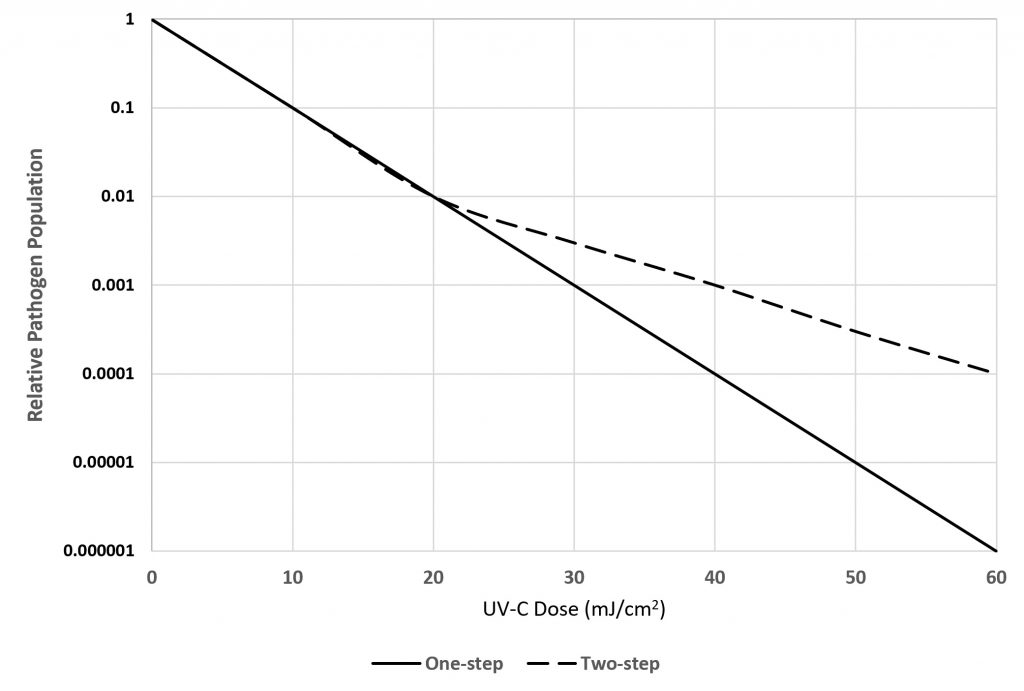
The third complication is that once D90 disinfection has been achieved, it is often the case that the surviving 10 percent of the population is an order of magnitude more resistant to UV irradiation. For example, if it takes 10 mJ/cm2 to achieve D90 disinfection, it may take 40 mJ/cm2 to achieve D99 disinfection.. As shown in Figure 2, such pathogens are said to have a “two-step” rare constant for their susceptibility to UV-C radiation.
FIG. 2 – One-step versus two-step pathogen susceptibility to UV-C radiation.
A fourth complication is that when vegetative bacteria and fungi have been exposed to monochromatic UV radiation as a means of disinfection, subsequent exposure to visible light may enable the “killed” cells to repair their DNA and recolonize, thereby increasing their survival rate by 10 to 100 times, especially if the pathogens are suspended in water or present on surfaces in the presence of high relative humidity. (Viruses do not seem to have the complexity needed to effect self-repair of their DNA.) This may be a concern if, for example, the exposed surfaces of a hospital room are decontaminated with a mobile UV disinfection robot, but the room is flooded with direct sunlight thereafter.
A fifth and final complication is that once a surface has been infected with respiratory droplets, any bacteria may find sufficient resources to begin colonizing the surface. The surface may be continuously irradiated by, for example, UV‑C radiation from a microplasma emitter or UV-LED array, but if the irradiance is too low, the surface may not achieve even D90 decontamination, regardless of the exposure time.
Summary
There is admittedly a considerable amount of information here that extends beyond the bounds of ultraviolet radiation terminology. It is needed, however, to put the terminology used for decades by the ultraviolet disinfection community into context. Ultraviolet radiation is not visible light, and so the lighting community needs to both understand and respect the terminology when adopting UVGI system design practices for building safety and human health. If we do not use the correct terminology, we risk (and deserve) a plague of apostilbs, brils, lamberts, skots and other annoyances later down the road.
Lighting designers will be familiar with the illuminance of a planar surface, which is measured in lumens per square meter (or foot). The irradiance of a planar surface by a germicidal radiation source is conceptually the same, except that it is measured in watts per square meter (W/m2). Most designers, however, will not be familiar with the concept of the spherical irradiance (or equivalently, fluence rate) of a point in space. This is an essential metric for air disinfection by germicidal lamps, and so it needs to be understood.
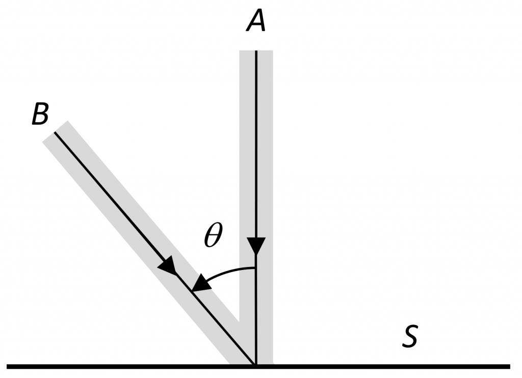
Figure 1 shows a parallel beam of UV radiation A with a cross-sectional power density of ΦA watts of power (flux) per square meter (W/m2) irradiating a surface oriented perpendicular to the beam. The beam can be made infinitesimally narrow, thereby irradiating a point on the surface, with irradiance EA = ΦA W/m2.
Figure 1. Surface irradiance.
Another parallel beam B with ΦB watts per square meter intersects the surface with incidence angle θ, the surface irradiance due to this beam being EB= (ΦBcos θ) W/m2. If we sum the contribution of beams from all possible directions above the surface of the plane, we obtain the surface irradiance ES. For germicidal applications, this irradiance is typically measured in microwatts per square centimeter (µW/cm2).
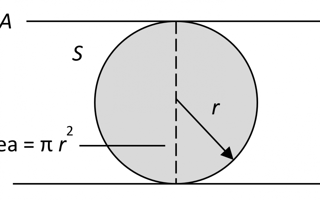
Irradiance is an appropriate metric for pathogens cultured on a plate, but aerosols are miniscule droplets of water suspended in air. If these droplets have been expelled by someone with an infectious respiratory disease such as tuberculosis or influenza, they may contain bacteria or viruses. These pathogens can be inactivated by UV radiation, but there is no planar surface that is being irradiated, and so the concept of irradiance does not apply.
Figure 2 represents the droplet as a transparent sphere S that does not absorb or refract UV radiation. The sphere has radius r, and so has a cross-sectional area π r2. If this sphere is irradiated by a beam A with cross-sectional power density ΦA W/m2, its irradiance is the same as the beam power density (again typically measured in µW/cm2). The diameter of the sphere can be made infinitesimally small, yielding an elementary sphere – a point in space – while the irradiance remains constant.
Figure 2. Spherical irradiance.
If we sum the contribution of beams from all possible directions of an imaginary sphere surrounding the point, we obtain the spherical irradiance of the point in space. This is also referred to as the fluence rate, which, when multiplied by the exposure time (and assuming a constant rate), yields the fluence of the droplet (typically measured in millijoules per square centimeter, mJ/cm2). If the fluence rate varies (as when the droplet moves through a three-dimensional UV radiation field), the fluence is the sum of the fluence rates multiplied by the chosen time intervals, which may vary from milliseconds to hours.
Finally, the term germicidal dose applies to both the irradiance of surfaces and the spherical irradiance of aerosols, but the terms fluence and fluence rate apply only to aerosols.
About Those Standards …
The concept of spherical irradiance may be simple to understand, but only if you ignore the official definitions. The CIE International Lighting Vocabulary has taken the approach that more is better, offering a choice of:
Section
Definition
17-742
Luminous spherical exposure (see 17-1028)
17-1244
Spherical illuminance (see 17-1245)
Photometric Quantities
Section
Definition
17-454
Fluence (see 17-1028)
17-455
Fluence rate (see 17-1245)
17-1023
Radiant fluence (see 17-1028)
17-1024
Radiant fluence rate (see 17-1245)
17-1028
Radiant spherical exposure
17-1245
Spherical irradiance
Radiometric Quantities
Section
Definition
17-883
Photobiological fluence (see 17-1028)
17-884
Photobiological fluence rate (see 17-1245)
Photobiological Quantities
Section
Definition
17-925
Photon fluence (see 17-1028)
17-926
Photon fluence rate (see 17-1245)
17-933
Photon spherical exposure (see 17-1028)
17-934
Photon spherical irradiance (see 17-1245)
Photon Quantities
In other words, spherical irradiance (or fluence rate) and fluence with different spectral weightings such as V(λ) for luminous quantities and photobiological action spectra, including photon quantities for horticultural applications.
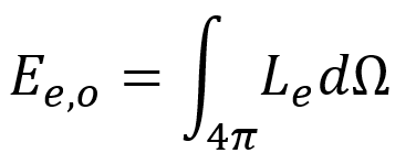
Trying to understand the CIE definition of fluence rate can be a challenge:
Quantity defined by the formula:
where dΩ is the solid angle of each elementary beam passing through the given point and Le its radiance at that point.
This makes sense if you think of an “elementary beam” as being an infinitesimally narrow cone (which makes it equivalent to the infinitesimally narrow beam discussed above), but there is the unnecessary complication of defining the beam radiance in terms of point sources. It is conceptually much easier to begin with parallel beams that can be made infinitesimally narrow.
ANSI/IES RP-16-17, Nomenclature and Definitions, on the other hand, is decidedly more spartan:
Luminous fluence
Luminous fluence rate
Radiant fluence
Radiant fluence rate
Spectral radiant fluence
Spectral radiant fluence rate
In addition to being spartan, the IES definitions are both clear and concise:
Fluence rate: The omnidirectional radiant flux externally incident on an elementary sphere about the point, per cross-sectional area of the sphere.
Fluence: The omnidirectional radiant energy externally incident on an elementary sphere about the point, per cross-sectional area of the sphere.
The advantage of these definitions is that they make no mention of radiance or solid angles.
Measuring Spherical Irradiance
CIE-ILV 17-1245, Spherical Irradiance, includes this helpful note:
This is the appropriate radiometric quantity for describing a dose rate for a photobiological or photochemical effect in a scattering medium (e.g., light in skin). It is also the appropriate quantity for describing the irradiation of microorganisms. It is frequently used incorrectly as a substitute for irradiance in some publications.
This leads to the obvious question: how do you measure spherical irradiance?
One approach is to use a spherical actinometer, an instrument that consist of a hollow quartz sphere measuring a centimeter or so in diameter that is filled with a solution of ferrioxalate, persulfate, or iodide/iodate; their transmittance after exposure is linearly proportional to the UV-C fluence (e.g., Kowalski 2009).
Another option is to use a radiometer with a “scalar irradiance” (yet another synonym for fluence rate) collector and a narrowband ultraviolet filter, such as the AMOUR radiometer manufactured by Biospherical Instruments (San Diego, CA). However, such an instrument has a “blind spot” of approximately 80 degrees where the spherical Teflon collector is mounted on its connector shaft.
UPDATE 20/08/28 – The manufacturer has stated that their AMOUR radiometer is not capable of measuring UV-C radiation due to excess absorption by the Teflon collector.
It may be difficult to measure spherical irradiance, but it is possible to predict its three-dimensional distribution in space using modified lighting design and analysis software, including contributions of direct radiation from UV-C sources and interreflections from surfaces. However, as Pierre de Fermat said regarding his Last Theorem, “I have discovered a truly remarkable theorem … which this margin is too small to contain.” More details to follow …
Acknowledgements
Thanks to Dawn DeGrazio of the Illuminating Engineering Society for her review and comments.
References
Kowalski, W. 2009. Ultraviolet Germicidal Irradiation Handbook: UVGI for Air and Surface Disinfection. Heidelberg, Germany: Springer.
There has been some discussion online and in presentations recently about the issue of photosynthetic photon flux. The argument goes as follows:
Photosynthetically Active Radiation (PAR) is somewhat arbitrarily defined as optical radiation within the spectral range of 400 nm to 700 nm.
Exposing plants to far-red radiation (defined as 700 nm to 800 nm) results in an increase in the rate of photosynthesis – the Emerson effect that was first noted in 1957 and confirmed by recent research.
Many horticultural luminaire manufacturers are now including far-red (725 nm) LEDs in their products.
The photosynthetic photon efficacy (PPE) of these luminaires is penalized by the definition of PAR because the far-red radiation is not taken into consideration.
The definition of PAR therefore needs to be changed to allow fair comparison of these products.
The one-word answer to this argument is … no.
Photosynthetic Photon Efficacy
ANSI/ASABE S640 JUL2017, Quantities and Units of Electromagnetic Radiation for Plants (Photosynthetic Organisms): defines Photosynthetic Photon Efficacy (PPE) as:
The photosynthetic photon efficacy (Kp) is the photosynthetic photon flux divided by input electric power. The unit is micromoles per second per electric watt (μmol × s-1 × We-1), or micromoles per joule (μmol × J-1).
Ignoring the technical jargon, the key point here is micromolesof photons. Photosynthesis occurs when a photon is absorbed by a photopigment (primarily chlorophyll A or B). In accordance with the Stark-Einstein law (aka the second law of photochemistry), one photon initiates one chemical reaction, regardless of the photon’s wavelength. We must therefore count the number of photons per second (measured in micromoles per second) rather than lumens or radiant watts for horticultural purposes.
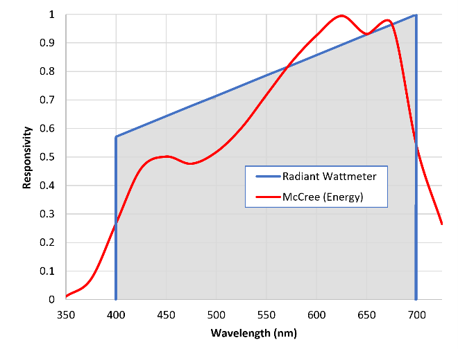
Referring to FIG. 1, McCree (1972) measured the relationship between wavelength and photosynthesis to produce the averaged “McCree curve.” He also acknowledged the Stark-Einstein law, which accounts for the blue line between 400 nm and 700m. What this means is that we can ignore the spectral power distribution of any light source within the range of 400 nm to 700 nm. As long as we have a calibrated PAR (aka “quantum”) sensor – which is basically a radiant wattmeter with the spectral response shown in FIG. 1 – we can measure micromoles of photons per second.
Emerson Effect
In a recent paper, Zhen and Bugbee (2020) presented an excellent argument in favor of redefining photosynthetically active radiation to include the spectral range of 400 nm to 750 nm. The title of the paper even includes the phrase, “Implications for Redefining Photosynthetically Active Radiation.”
The authors are unquestionably correct; far-red photons in effect supercharge the process of photosynthesis, and must – not should, but must – be taken into consideration when defining photosynthetically active radiation.
This does not mean however that the PAR metric should be redefined. Quoting from the abstract of Zhen and Bugbee (2020): “Far-red alone minimally increased photosynthesis … far-red photons are equally efficient at driving canopy photosynthesis when acting synergistically with traditionally defined photosynthetic photons.” In other words, if we assume a spectral range of 400 nm to 750 nm, we cannot unambiguously measure the photosynthetic photon efficacy of a light source without knowing its spectral power distribution. That is, without knowledge of the entire spectral power distribution within this range, we cannot predict the rate of photosynthesis.
Figure 1 – McCree curve and PAR sensor response.
To rephrase the issue, the current definition of PAR assumes that the photosynthesis rate of higher plants is linear with respect to incident radiation within the spectral range of 400 nm to 700 nm. There are obviously minimum and maximum irradiance limits to where this assumption applies, but it is necessary in order for the concept of PAR and hence PPE to have any meaning.
The Emerson effect violates this assumption by making the photosynthesis rate nonlinear – add far-red radiation beyond 700 nm and you will change the rate in the manner that depends on the spectral power distribution of the PAR radiation.
This issue does not concern just horticultural luminaires with 725 nm far-red LEDs. Most red-emitting phosphors used in white-light LEDs and red-emitting phosphor-coated LEDs (e.g., Figure 2) have significant emissions in the far-red, and so may invoke the Emerson effect.
Regardless, it remains that the definition of PAR cannot redefined. It is not a matter of penalizing horticultural luminaires with far-red emissions, but of simply having a metric that makes sense.
Photomorphological Considerations
There is an additional complication with far-red radiation. Horticultural luminaires including far-red LEDs typically employ 660 nm red and 725 nm far-red LEDs. These wavelengths correspond nicely with the peak absorptances of the Pr and Pfr isoforms of phytochrome, a plant photoreceptor that is responsible for plant morphology from seed germination to leaf senescence, shade avoidance, and circadian rhythms. By offering luminaires with fixed red to far-red (R:FR) ratios, luminaire manufacturers have only begun to explore the horticultural possibilities of far-red radiation.
Does the current definition of Photosynthetically Active Radiation and hence Photosynthetic Photon Efficacy disadvantage horticultural luminaire manufacturers who include far-red LEDs in their products? In one sense, the answer is yes. However, this is a very narrow view of the issue that focuses on a single metric. The goal should be to educate the customer that despite a possibly lower PPE value for the product, the far-red radiation represents a value-added feature.
References
ANSI/ASABE S640 JUL2017, Quantities and Units of Electromagnetic Radiation for Plants (Photosynthetic Organisms). St. Joseph, MI: American Society of Agricultural and Biological Engineers.
McCree, K. J. 1972a. “The Action Spectrum, Absorptance and Quantum Yield of Photosynthesis in Crop Plants,” Agricultural and Forest Meteorology 9:191-216.
Zhen, S., and B. Bugbee. 2020. “Far-red Photons Have Equivalent Efficiency to Traditional Photosynthetic Photons: Implications for Redefining Photosynthetically Active Radiation,” Plant Cell Environ. 2020:1-14. DOI: 10.1111/pce.1370.
Germicidal lamps emitting ultraviolet-C (UV-C) radiation have been in use since the 1930s (Wells and Wells 1936). These are most commonly low-pressure mercury-vapor discharge lamps, which are basically fluorescent lamps without a phosphor coating and fused quartz rather than borosilicate glass bulbs. They emit monochromatic radiation mostly at 254 nm, a wavelength that is very effective in disrupting the DNA of viruses, bacteria, and other pathogens.
The photobiological risks of these germicidal lamps are well-known: exposure to UV-C radiation can result in photokeratitis (“snow blindness”), photoconjunctivitis (“pink eye”), and erythema (sunburn). These medical conditions typically only last for a few days, but they can be quite painful. Unlike UV-B radiation (280 nm to 315 nm), UV-C radiation is much less likely to cause long-term cellular damage leading to skin cancer,
More recent germicidal light sources include UV-C light-emitting diodes and pulsed xenon discharge lamps, but there is a newcomer on the block that has gained considerable media attention: far-ultraviolet excimer lamps. Recent medical studies have indicated that, unlike 254 nm radiation, the 207 nm and 222-nm “far-UV” radiation emitted by excimer lamps is likely harmless (e.g., Buonanno et al. 2017, Welch et al. 2018). Excimer lamps have the same germicidal properties as mercury-vapor discharge lamps, but the shorter wavelength radiation cannot penetrate deeply enough into the outermost cells of the eyes and skin to disrupt their DNA.
This leads to the exciting thought that we may be able to design UV-C germicidal systems using far-UV excimer lamps. Unlike mercury-vapor lamps and UV-C LEDs, there does not appear to be any significant photobiological risk (if their residual UV-C emissions are blocked), and so they could be deployed in direct view of the room occupants while disinfecting both the air and contaminated surfaces with their radiation.
Indeed, there are already companies advertising such products, although they do not appear to be commercially available as yet. This does not stop us, however, from asking the question: what does it take to design a UV-C disinfection system using far-UV radiation?
Excimer Lamps
Excimer lamps consist of diatomic molecules that form a plasma when an electrical current passes through them. A combination of krypton and chlorine (KrCl) gases, for example, emits 222-nm radiation, while krypton and bromine emit 207-nm radiation.
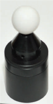
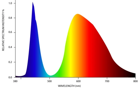
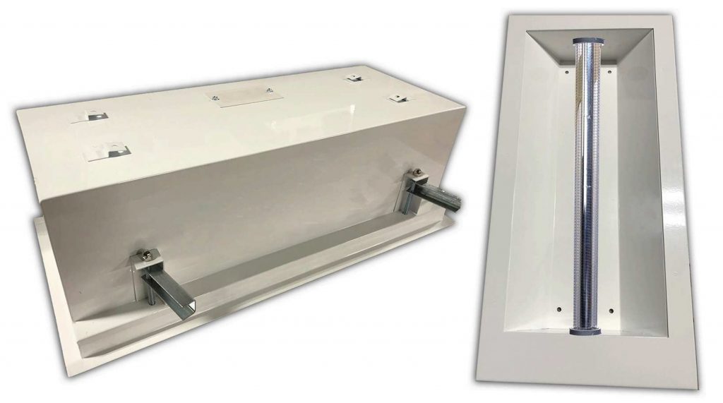
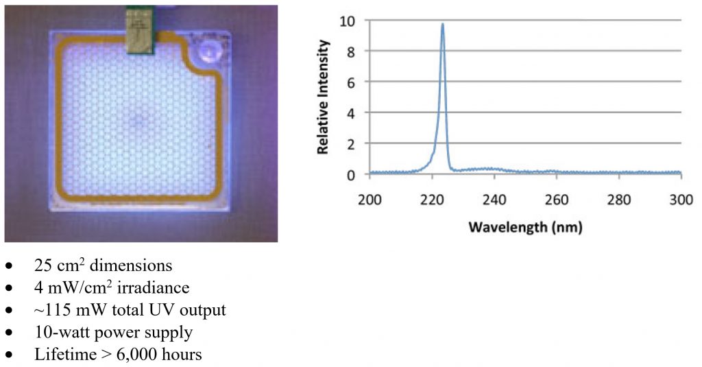
Companies such as Ushio and SterilRay manufacture excimer lamps and products for industrial and medical applications, but the lamps are typically comparable to fluorescent lamps in size and form factor (e.g., FIG. 1). However, one company – Eden Park Illumination – has adapted the technology formerly used in plasma television displays to produce thin microplasma lamps intended for general illumination purposes. One of their evaluation products is of particular interest, as it generates nearly monochromatic 222 nm UV-C radiation (FIG. 2).
The published specifications of this product are not particularly remarkable, but they are useful in that they enable us to evaluate the usefulness of this technology for germicidal applications. Eden Park states that they have achieved a maximum irradiance greater than 25 μW/cm2 in the laboratory, but this is presumably with a much shorter lifetime. (The criterion for lifetime is not defined, but presumably refers to UV-C output power depreciation over time.)
Germicidal Dose
A key characteristic of germicidal lamps of any sort is the UV-C radiation dose (irradiance multiplied by exposure time), expressed in millijoules per square centimeter (mJ/cm2). The required dose depends on both the pathogen species to be eliminated and the desired degree of reduction. For example, eliminating 90 percent of Escherichia coli O157:H7, the bacterium that causes sometimes fatal food poisoning, requires 1.5 mJ/cm2; doubling the dose eliminates 99 percent, tripling eliminates 99.9%, and so forth. This is referred to as log10 (“log-ten”), or more commonly “log,” reduction:
Log10 reduction
Percent pathogen elimination
1
90%
2
99%
3
99.9%
4
99.99%
5
99.999%
Table 1 – Log10 pathogen reduction.
The International Ultraviolet Association publishes a compilation of dose requirements for many different pathogens, but viruses on average require a dose of about 20 mJ/cm2 for 90 percent reduction when directly exposed to the UV-C radiation (IUVA undated). Most of the studies referenced in the compilation consider 254 nm radiation from low-pressure mercury vapor lamps, but the required dose from 207-nm and 222-nm excimer lamps should be comparable.
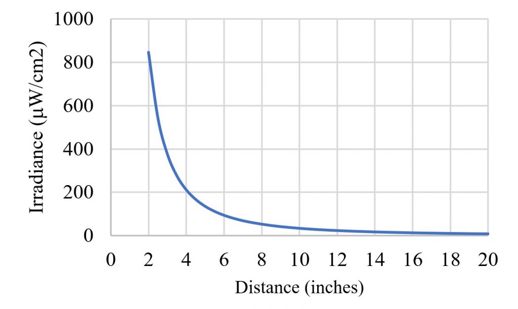
The goal, of course, is to provide sufficient UV-C irradiance that the desired log reduction is achieved within an allotted time. With this in mind, it is useful to calculate the expected irradiance versus distance for the 222-nm microplasma excimer lamp (FIG. 3).
Figure 3 – Irradiance from 25 cm2 microplasma lamp versus distance.
It should be noted that the excimer lamp is an area source, and so the inverse square law does not apply in the near-field, or a distance of less than 10 inches. It was also assumed that the lamp has a Lambertian (i.e., cosine) radiant intensity distribution.
This plot is useful in that the irradiance versus distance values will give us a sanity check for the next phase of the design. For example, if we have a requirement for 90 percent virus reduction in 20 seconds, we would need an irradiance of 1,000 μW/cm2, requiring a distance of less than two inches from the lamp. Even if the lamp produced the maximum reported output of 25 μW/cm2, the maximum distance would still be less than five inches.
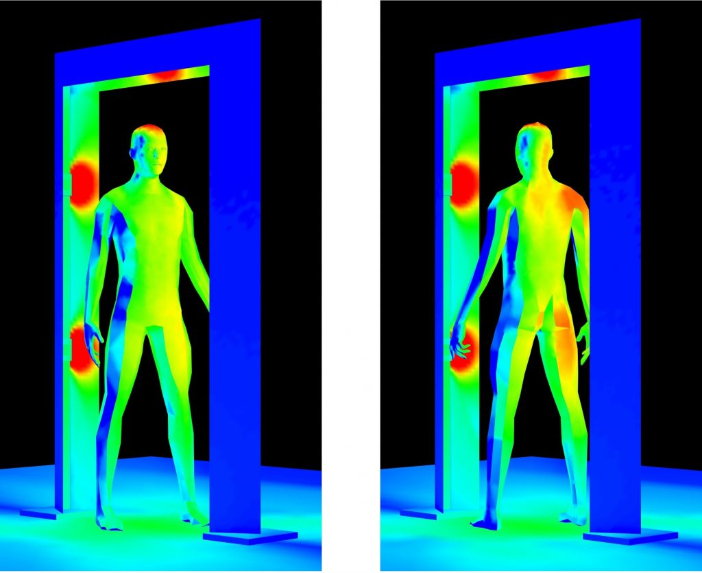
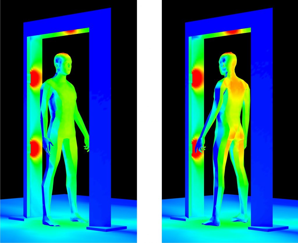
Doorway Disinfection
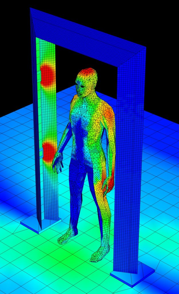
A possible design of obvious interest is to place the excimer lamps in the frame of a doorway or entry portal, much like an airport security scanner. Anyone passing through the doorway can – in theory – be disinfected.
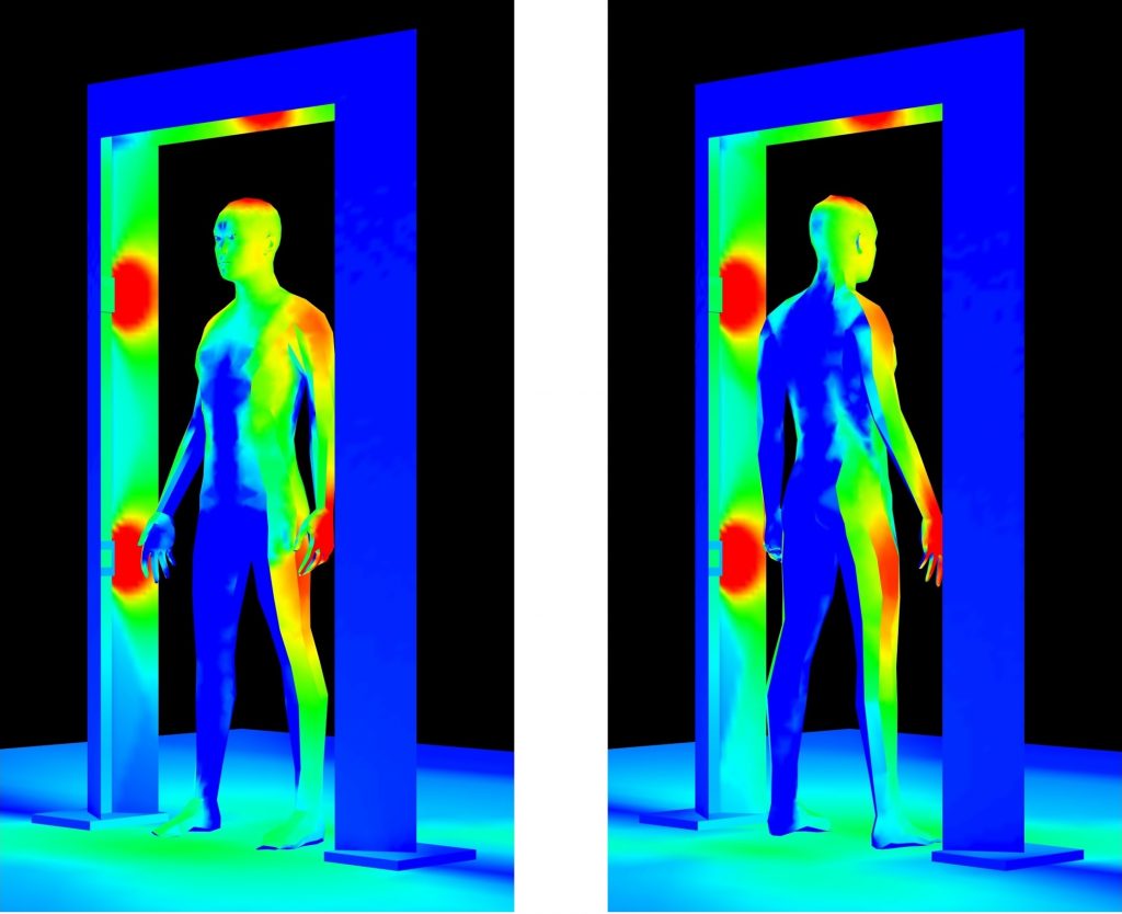
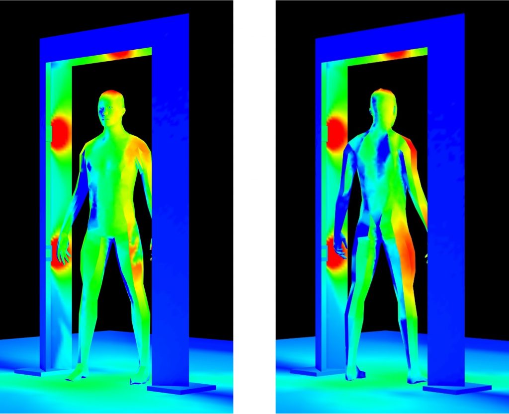
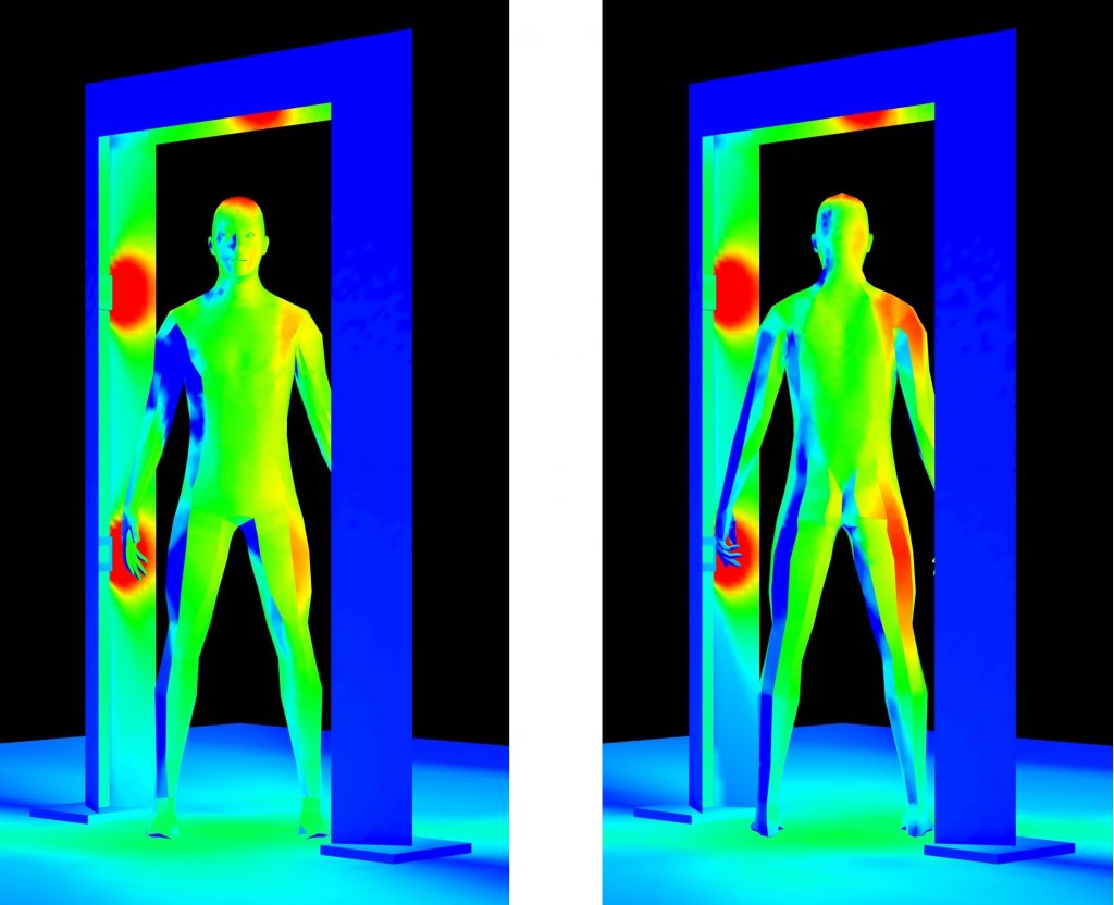
In theory … the goal here is not to design a specific disinfection system, but to consider the factors that go into its design. What we learn from this exercise can be used to guide engineering design for commercially-realizable disinfection systems. Conceptually then, we want a system wherein a person walks into the doorway, does a complete 360-degree turn, then continues on after being disinfected by the (presumably) safe 222-nm UV-C radiation.
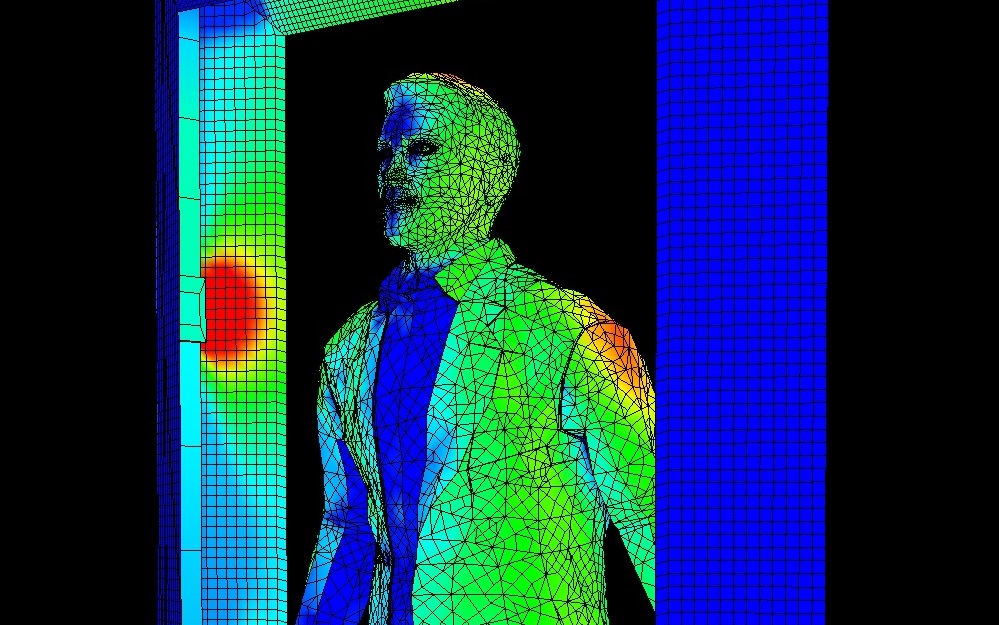
The doorway shown in Figure 4 has an opening 50 inches wide by 80 inches high. It has four microplasma lamps mounted at 30 inches and 60 inches above the floor, and a fifth lamp mounted overhead. The pseudocolor heat map shows the far-UV irradiance due to these lamps.
Figure 4 – Proposed doorway disinfection system.
It is in general difficult to obtain UV-C reflectance data for most materials. A report published four decades ago summarized the results of studies done between the 1920s and 1940s, but very little information has been published since then (Ullrich and Evans 1976). Still, there is sufficient data available to model the system shown in Figure 3 (Cader and Jankowski 1998 and Nagy 1964):
The reflectance of human skin to UV-C radiation is less than one percent.
The reflectance of oil-based paints is 5 to 10 percent.
The reflectance of white cotton is about 30 percent.
The reflectance of etched aluminum is 88 percent.
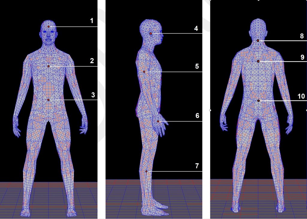
Given this, we can place a virtual mannequin inside the doorway, rotate it through 90 degrees, and measure the predicted far-UV irradiance at selected target points (FIG. 5). The results are shown in Table 2 and Figure 6.
The design certainly has a few shortcomings, although we could have expected them from Figure 2. If the goal is to achieve a 90% reduction in pathogens (99.9% is preferable), we would need a far-UV dose of 20 mJ/cm2. With a minimum irradiance value of 2.55 μW/cm2, we would need therefore an exposure time of over two hours!
It gets worse, however. The UV-C doses required to achieve a given reduction in pathogens are determined by irradiating cultures in Petri dishes and test tubes. Disinfecting surfaces in the real world typically requires larger – sometimes much larger – doses. Increasing the number of excimer lamps would of course increase the average irradiance, but this approach has its limits. The American Conference of Governmental Industrial Hygienists recommends a maximum exposure of 3.0 mJ/cm2 of broadband (200 nm – 315 nm) ultraviolet radiation per 8-hour workday, and specifies a spectral weighting function to assess ultraviolet hazards for skin and eye (ACGIH 2013). For 222-nm radiation, the weight factor is 0.12, meaning that a maximum exposure of 25 mJ/cm2 is recommended. At 1,000 μW/cm2, a 30-second exposure would exceed the recommended daily limit. This would be a concern not only for the person being irradiated, but also for any service personnel standing near the doorway for extended periods of time.
It must be acknowledged that the ACGIH spectral weighting function is based on medical studies of photokeratitis and erythema performed prior to 1991, and so do not take recent studies of far-UV exposure into account. Nevertheless, until such time as the spectral weighting function is revised, it remains a standard for UV-C exposure limits.
One issue that will need to be addressed is that krypton-chlorine lamps emit about three percent of their radiation in the region of 230 to 260 nm, as can be seen in Figure 2. There is evidence that far-UV exposure can cause erythema in persons with phototypes I and II skin, and also patients taking photosensitizing drugs (e.g., Woods 2015 and Saadati 2016). This reaction is likely due to the residual UV-C radiation, but it can presumably be blocked by suitably doped fused quartz filters.
There is also a fundamental flaw in this design: the SARS-CoV-2 virus that causes COVID-19 appears to be spread primarily through aerosols generated by coughing, sneezing, and even talking. Even if the doorway disinfection system were capable of properly disinfecting surfaces, it would have no effect on an infected person walking through it.
Security Theater
The unfortunate conclusion is that using microplasma excimer lamps in doorway disinfection systems fails by several orders of magnitude. From an engineering perspective, this is an undesirable outcome. However, we should not be surprised to see such systems in common use in the near future. We have lived with airport security full-body scanners for years. It is widely acknowledged these devices are an example of security theater – the practice of investing in countermeasures intended to provide the feeling of improved security while doing nothing or little to achieve it. Doorway disinfection systems – with the associated annoyance of having to pause for 20 seconds or so before entering a building – may become just as common in our daily lives.
More Research Required
None of the above should be construed as a criticism of far-UV excimer lamps for room disinfection. Based on the evidence to date, they appear to be safer and equally as effective as low-pressure mercury-vapor discharge lamps for upper-room air disinfection and surface disinfection in unoccupied spaces. However, more research is required to determine whether the existing ACGIH dose recommendations for far-UV radiation can be exceeded.
References
ACGIH. 2013. Ultraviolet Radiation: TLV(R) Physical Agents 7th Edition Documentation. American Conference of Governmental Industrial Hygienists.
Buonanno, M., et al. 2017. “Germicidal Efficacy and Mammalian Skin Safety of 222-nm UV Light,” Radiation Research 187(4):483-491. DOI: 0.1667/RR0010CC.1.
Cader, A., and J. Jankowski. 1998. “Reflection of Ultraviolet Radiation from Human Skin Types,” Health Physics 74(2):169-172.
Nagy, R. 1964. “Application and Measurement of Ultraviolet Radiation,” American Industrial Hygiene Association Journal 25:274-281.
Narla, S., et al. 2020. “The Importance of the Minimum Dosage Necessary for UV-C Decontamination of N95 Respirators During the COVID-19 Pandemic,” Photodermatology, Photoimmunology & Photomedicine. DOI: 10.1111/phpp.12562.
Saadati, S. 2016. Study of Ultraviolet C Light Penetration and Damage in Skin. Department of Radiophysics, Sahlgrenska University Hospital. Gothenburg, Sweden.
Ullrich, O. A., and R. M. Evans. 1976. Ultraviolet Reflectance of Paints. American Welding Society.
Welch, D., et al. 2018. “Far-UVC Light: A New Tool to Control the Spread of Airborne-Mediated Microbial Diseases,” Scientific Reports 8:2752. DOI: 10.1038/s41598-018-21058-w.
Wells, W., and M. Wells. 1936. “Air-borne Infection,” J. American Medical Association 107:1069:1703.
Woods J. A., et al. 2015. “The Effect of 222-nm UVC Phototesting on Healthy Volunteer Skin: A Pilot Study,” Photodermatology, Photoimmunology & Photomedicine 31(3):159- 166.
This website uses cookies (mostly chocolate chip) to improve your experience. We'll assume you're ok with this, but you can opt-out if you wish. Cookie settingsACCEPT
Cookies Policy
Privacy Overview
This website uses cookies to improve your experience while you navigate through the website. Out of these cookies, the cookies that are categorized as necessary are stored on your browser as they are essential for the working of basic functionalities of the website. We also use third-party cookies that help us analyze and understand how you use this website. These cookies will be stored in your browser only with your consent. You also have the option to opt-out of these cookies. But opting out of some of these cookies may have an effect on your browsing experience.
Necessary cookies are absolutely essential for the website to function properly. This category only includes cookies that ensures basic functionalities and security features of the website. These cookies do not store any personal information.
Any cookies that may not be particularly necessary for the website to function and is used specifically to collect user personal data via analytics, ads, other embedded contents are termed as non-necessary cookies. It is mandatory to procure user consent prior to running these cookies on your website.